Epidemiology, Infectious Diseases, Parasites, Pathogen of the Month, tick-borne diseases

Babesiosis: A Rare But Deadly Blood Infection Spread by Ticks

Author Chandana Balasubramanian , 06-Jul-2022

Babesiosis in humans is a rare, infectious, tick-borne disease caused by a group of single-celled parasites belonging to the genus Babesia. The disease affects and destroys red blood cells, leading to a special type of anemia known as hemolytic anemia, characterized by jaundice and darkened urine.


As tick-borne illnesses go, babesiosis has stayed in the shadows compared to its more well-known “cousin” Lyme disease. But babesiosis is now an emerging infectious disease in Southeastern, Central, and Northeastern Europe as tick populations grow rapidly and spread to non-endemic regions. It’s time to start paying more attention to this potentially life-threatening illness [1].


Babesiosis goes by many other names, including piroplasmosis, tick fever, Texas fever, or Nantucket fever, and is spread by hard-bodied (ixodid) ticks — the very same that spread Lyme disease. An issue with diagnosing babesiosis is that the symptoms are very similar to ehrlichiosis, another tick-borne disease, and mosquito-borne malaria. As a result, babesiosis cases may be under-reported in countries where malaria is endemic [2].


History of This Rare Disease


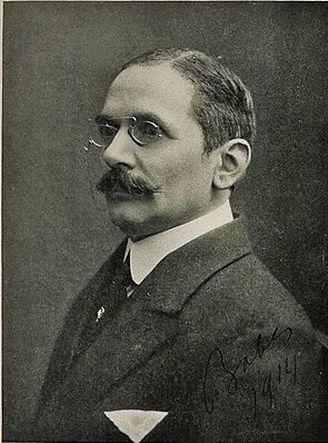
The parasite Babesia was named after Romanian scientist Victor Babes, who was the first to discover babesiosis pathogens in the red blood cells of cattle in the year 1888. Later in 1892, he observed similar organisms in sheep blood [3]. Babes was a prominent physician and one of the first few bacteriologists in the world. He is also considered one of the founders of modern microbiology and the second radiologist in the world (after Louis Pasteur) to introduce rabies vaccines into Romania.

Victor Babes Image from KUMC

In 1893, Smith and Kilborne in the United States also identified similar organisms in cattle blood that were later called Babesia bigemina and found to cause cattle fever. Over time, more than 100 babesia species have been identified in animals [4].

The first case of human babesiosis was identified in 1957 in a farmer in Croatia (at the time, a part of Yugoslavia). The farmer presented with fever, anemia, and hemoglobinuria. At the time, the causative agent was unknown but was later found to be B.divergens [4].

Since then, several other species of babesia causing infections in humans have been identified. Of these, B. microti, B. duncani, and B. venatorum are the parasites responsible for the majority of cases in humans [5].

B. microti was first identified in an immunocompromised person in the US in 1969 [5]. B. duncani was also identified in the United States in 1991 [6]. In 2003, B. venatorum was identified in Italy [2,4].

Together, these four babesia species are confirmed to be zoonotic pathogens [2].


Epidemiology


Though there are several species of babesia, only a few are known to cause human babesiosis. They include B. divergens, B. duncani, B. microti, and B. venatorum [5]. The disease caused by these species can be severe, mild, or even asymptomatic and varies depending on the infected individual’s health. It can be life-threatening for those with poor immunity, those who have had their spleen removed, or those with underlying health problems like liver or kidney disease [7].

In 2011, around 1,124 human babesiosis cases were reported in the US. Since then, the number of cases reported to the CDC has gradually increased. The highest number of cases in the US were reported in the year 2019, with a total of 2,418 cases. This is an 11% increase from the number of cases reported during 2018 and a 115% increase from those reported in 2011 [7].

United States. Babesiosis, cases (Rates per 100,000) Chart by GIDEON

B. microti was the causative agent for most of these cases [5,8]. B. microti infections were found to have a mortality rate of about 5% [9]. In the United States, the B. microti parasite is endemic to the mid-western (which includes the states of Minnesota and Wisconsin) and the northeastern regions (which includes the states of New Jersey, New York, Connecticut, Massachusetts, and Rhode Island). This is because of a higher prevalence of white-footed mice, one of the primary hosts for the ticks that transmit this infection [8]. Australia, Japan, and Taiwan have also reported a few human babesiosis cases caused by B. microti [10].

Compared to B. microti, infections caused by B. divergens are quite rare [8]. However, B.divergens is the main source of human babesiosis infections in Europe. As of 2003, 31 cases had been reported in Europe, out of which nine turned out to be fatal [11]. As of 2021, out of 51 cases documented in Europe, 35 were found to have been caused by B. divergens [12].

Most of these cases were reported in countries with extensive cattle industries, such as the UK, France, and Ireland. This is because cattle are one of the primary hosts for the ticks that carry the infection [10].

Infections caused by B. duncani and B. venatorum are significantly less than B. microti and B. divergens. So far, all of the nine cases of B. duncani reported were from the west coast of the United States [13]. B. venatorum infection cases were reported in Austria, Germany, and Italy [4]. The endemic range of B. venatorum also extends to Asia, Africa, and South America, and the disease caused by this species is not fatal [4, 13].


How It Is Spread and the Importance of Blood


Babesiosis infection cannot be transmitted from human to human except through blood transfusions, but the infection can spread to humans through:

  • Ticks: The most common mode of transmission of human babesiosis infection is through tick bites. People who are actively engaged in outdoor activities in endemic regions are susceptible to such infections. Rodents (usually white-footed mice), rabbits, deer, and cattle [14].
  • Blood transfusions: Babesiosis can also be transmitted through blood transfusions. This can happen when a person receives blood from a donor infected with Babesia but an asymptomatic donor [10,14].
  • Congenital transmission: People who are pregnant can pass their babesiosis infection to their fetus during pregnancy [14].


Biology of the Disease


Once Babesia enters the human body, they begin to target and invade the red blood cells (RBCs). In the RBCs, the parasites begin their cycle of maturation and growth. Once their growth cycle is completed, they exit the host cells (RBCs) while destroying them in the process. They are then released into the bloodstream after each cycle, and then they invade other uninfected RBCs. This is how the cycle of infection is repeated [8].


Diagnosis


Treatment for babesiosis usually begins only if the infected person is strongly suspected to be carrying the infection. Here’s what to consider when evaluating whether a person is suffering from babesiosis or not. Has the infected individual:

  • Recently traveled to or lived in a region endemic to babesiosis?
  • Spent a lot of time outdoors in an endemic region during the summer or the beginning of autumn?
  • Had a blood transfusion in the last six months?


If the answer to any of the questions above is ‘yes,’ then, the person can be strongly suspected to be infected with babesiosis [4,5]. Lab tests are required to confirm a babesiosis diagnosis. Giemsa or Wright staining of thin blood smears helps detect the presence of babesia parasites in the blood [5,8]. If the test result is positive, then, the patient may be given antimicrobial therapy [5]. In mild cases, however, it can be difficult to detect these parasites; serial blood smears may be needed depending on disease severity [5,8]. Assays based on PCR can also be done for a more accurate diagnosis. Similar to blood smears, depending on the severity of illness, PCRs can be repeated if the results are negative [5].


Symptoms


The incubation period for babesiosis varies based on the mode of transmission. If a tick bites a person, it can take one to four weeks for symptoms to appear. If an individual got a babesiosis infection from a transfusion with contaminated blood, the incubation period could vary from one to nine weeks.


In most cases, people remain asymptomatic (50% of children and 25% of adults), but some experience mild to severe symptoms. Fatigue, malaise, muscle pain, and persistent or intermittent pattern of fever, mark the onset of the disease. They will be followed by other symptoms like:

  • Chills
  • Cough
  • Dark-colored urine
  • Depression
  • Diarrhea
  • Difficulty in breathing
  • Eating disorder
  • Emotional lability – a psychological condition characterized by mood swings.
  • Abdominal pain
  • Headache
  • Joint pain
  • Redness of eyes
  • Sore throat
  • Sweats
  • Vomiting


Other clinical manifestations include:

  • Liver enlargement
  • Throat infection
  • Spleen enlargement
  • Jaundice
  • Bleeding and other issues with the retina [4,15]


Babesiosis symptoms can be similar to other diseases, which makes it very important to perform a thorough differential diagnosis at point-of-care. For example, ehrlichiosis is a tick-borne illness that infects white blood cells but shares some common symptoms with babesiosis, like fever, headaches, malaise, muscle pain, nausea, vomiting, cough, etc. [16].

Additionally, babesiosis is pathologically similar to malaria in many ways: both diseases are caused by single-cell eukaryotes, and both pathogens invade and multiply within the red blood cells of the host [17]. The two diseases share so many common symptoms and can be indistinguishable without proper diagnosis [8]. Because the two diseases are similar, the actual number of babesiosis cases is largely underestimated in regions where malaria is endemic [2].


Treatment


There are two medical treatments that are commonly administered to cure babesiosis: A combination of atovaquone and azithromycin, and a combination of clindamycin and quinine [4]. However, treatment varies depending on how severe the illness is and the type of babesiosis species causing the infection [4,8].


Mild illness: For immunocompetent people with mild babesia microti infection, antimicrobial medicines such as atovaquone and azithromycin are administered for seven to ten days. For those infected with babesia divergens or babesia venatorum, clindamycin is administered for seven to ten days. Quinidine or quinine is administered to treat mild human babesiosis cases caused by babesia duncani [8].


Severe illness: For severe illness, clindamycin is administered intravenously in combination with quinine for seven to ten days. In the case of a life-threatening infection caused by b. divergens, an exchange blood transfusion is performed. It is an effective way to decrease the load of parasites and remove toxic byproducts caused by the infection. Exchange blood transfusion is also done for patients suffering from severe babesiosis with a parasitemia (parasite load) of 10% and above [4,8].


Asymptomatic cases: No treatment is needed for asymptomatic patients even if they test positive for babesiosis. However, if parasitemia in the blood persists for over three months, antimicrobial treatment has to be given. Immunocompromised patients who are asymptomatic but have persistent parasitemia should have blood smears performed periodically until they are totally free from the infection [4].


Prevention


There is no vaccine for babesiosis [15]. The best way to prevent the disease is through preventive measures:

  • Raise public awareness about the symptoms of tick-borne diseases [5].
  • Avoid places with a high deer population since ticks usually feed on large animals like deer. Tick density is high during the summer and early autumn seasons, but tick infections can be found all year round [4,5].
  • Immunocompromised persons living in regions endemic to large tick populations should be advised to stay away from forests, woodlands, or other places where ticks are found in large numbers [4,5,15].
  • People visiting forests or woodlands should ensure they cover their bodies completely with protective clothing to prevent exposure to ticks [4,5,15].
  • Make it a practice to check for ticks attached to the body after any outdoor activity. Ticks must be removed with tweezers, not with the hands [4].
  • Tick repellents designed to prevent tick bites can be applied to areas of the body that cannot be covered by clothing. There are tick repellents that can be sprayed on clothes as well [4,5,15].
  • Certain pesticides can be sprayed in places with a high tick density to kill ticks that feed on mice and help contain the local tick population [4,5]. 
  • Drains, walls, and compounds can be sealed to prevent mice from entering the property [4].
  • Make sure that the land surrounding your home is not brushy or grassy. Mow regularly, if possible [4,5].
  • Before a blood transfusion, hospitals must screen a donor’s blood for any trace of babesia [15].


References

[1] A. Bajer et al., “Babesiosis in southeastern, central and northeastern Europe: An emerging and re-emerging tick-borne disease of humans and animals,” Microorganisms, vol. 10, no. 5, p. 945, 2022.

[2] E. Montero, J. Gray, C. A. Lobo, and L. M. González, “Babesia and Human Babesiosis,” Pathogens, vol. 11, no. 4, p. 399, Mar. 2022.

[3] L. T. S. Santos, “Generation of polyclonal and monoclonal antibodies against Babesia ovis protein extract (Doctoral dissertation),” Instituto de Higiene e Medicina Tropical, Universidade Nova de Lisboa, 2020.    

[4] E. Vannier, B. E. Gewurz, and P. J. Krause, “Human babesiosis,” Infect. Dis. Clin. North Am., vol. 22, no. 3, pp. 469–88, viii–ix, 2008.

[5] T. K. Chen and P. J. Krause, “Human Babesiosis,” in Clinical Infectious Disease, D. Schlossberg, Ed. Cambridge: Cambridge University Press, 2013, pp. 1381–1388.

[6] S. Wang et al., “Establishment of a transient and stable transfection system for Babesia duncani using a homologous recombination strategy,” Front. Cell. Infect. Microbiol., vol. 12, p. 844498, 2022.

[7] M. Swanson, “Surveillance for Babesiosis,” Centers for Disease Control and Prevention, 2021. [Online].   

[8] R. L. Ord and C. A. Lobo, “Human babesiosis: Pathogens, prevalence, diagnosis and treatment,” Curr. Clin. Microbiol. Rep., vol. 2, no. 4, pp. 173–181, 2015.

[9] M. J. Homer, I. Aguilar-Delfin, S. R. Telford 3rd, P. J. Krause, and D. H. Persing, “Babesiosis,” Clin. Microbiol. Rev., vol. 13, no. 3, pp. 451–469, 2000.

[10] J. S. Shah, N. S. Harris, I. R. Horowitz, and R. Horowitz, “Human Babesiosis and Ehrlichiosis -Current Status,” Igenex.com. [Online]

[11] A. Zintl, G. Mulcahy, H. E. Skerrett, S. M. Taylor, and J. S. Gray, “Babesia divergens, a bovine blood parasite of veterinary and zoonotic importance,” Clin. Microbiol. Rev., vol. 16, no. 4, pp. 622–636, 2003.

[12] A. Kumar, J. O’Bryan, and P. J. Krause, “The global emergence of human babesiosis,” Pathogens, vol. 10, no. 11, p. 1447, 2021.

[13] E. Vannier and P. J. Krause, “Update on babesiosis,” Interdiscip. Perspect. Infect. Dis., vol. 2009, p. 984568, 2009.

[14] CDC-Centers for Disease Control and Prevention, “CDC – Babesiosis – epidemiology & risk factors,” 2010.

[15] S. C. Parija, D. Kp, and H. Venugopal, “Diagnosis and management of human babesiosis,” Trop. Parasitol., vol. 5, no. 2, pp. 88–93, 2015.

[16] J. G. Schneider, B. S. Mt, and J. S. Dumler, “Clin Lab Sci 2009;22(1):3,” Ascls.org. [Online]

[17] A. S. Paul, C. K. Moreira, B. Elsworth, D. R. Allred, and M. T. Duraisingh, “Extensive shared chemosensitivity between malaria and babesiosis blood-stage parasites,” Antimicrob. Agents Chemother., vol. 60, no. 8, pp. 5059–5063, 2016.

Author
Chandana Balasubramanian

Chandana Balasubramanian is an experienced healthcare executive who writes on the intersection of healthcare and technology. She is the President of Global Insight Advisory Network, and has a Masters degree in Biomedical Engineering from the University of Wisconsin-Madison, USA.

Articles you won’t delete.
Delivered to your inbox weekly.