Bacteria, Infectious Diseases, Pathogen of the Month

Bartonellosis: Cat Scratch Disease, Carrion’s Disease, Trench Fever, and Bartonella Endocarditis

Author Chandana Balasubramanian , 22-Jun-2022

Bartonellosis is the term used to describe a group of infectious diseases caused by the bacteria Bartonella. These diseases include trench fever, Carrion’s disease (Oroya fever and Peruvian warts), and cat scratch disease, to name a few. They affect both animals and humans [1]. Bartonellosis diseases are zoonotic: they can be transmitted from domestic animals to humans.  Various insects, including fleas, body lice, and ticks, act as vectors and play an important role in transmission — between animals and between animals and humans. 


The occurrence of Bartonellosis is most commonly associated with areas of high population density and poor sanitation [2]. Species of Bartonella are fastidious, gram-negative bacteria. Let’s take a closer look at the most common Bartonella bacterial infections:

  • Cat Scratch Disease (CSD)
  • Carrion’s Disease 
  • Endocarditis 
  • Trench Fever 

Cat Scratch Disease (CSD)


Cat scratch disease is also known as cat scratch fever (CSD) or subacute regional lymphadenitis. It is the most common Bartonella infection. The disease is caused by the species Bartonella henselae and infects the lymph nodes near the sites of cat scratches, causing them to swell. It is a common source of swollen lymph nodes (lymphadenopathy) in children and teenagers. On occasion, flea or tick (arthropod) vectors can directly transmit CSD to humans [3]. 


History


Cat scratch disease was first described in 1931 by Dr. Robert Debré. The doctor observed regional lymph nodes swelling after cat scratches. Twenty years later, he published his report on CSD, which officially recognized the infection as a clinical condition [4]. However, B. henselae, the causative agent, was not identified until the 1990s through PCR amplification [5]. 

Epidemiology


CSD has a global incidence of 6.4 cases per 100,000 adults and 9.4 cases per 100,000 children aged between five to nine years [1]. Incidence varies from place to place and depends on the regional population of fleas responsible for the transmission of the infection. Places that are warm and humid usually have more fleas and therefore have a higher incidence than arid areas (including mountain regions), where the disease incidence is much lower. Children between the age group of five to nine years are most affected [6].

How is it Spread?


Cat scratch disease is transmitted when an infected cat bites or scratches an individual. It can also spread when an infected cat licks an open wound on a person [7]. According to the CDC, approximately 40% of cats carry CSD in their lifetime, even though many remain asymptomatic. Kittens that are younger than one year are more likely to carry and transmit this disease. 

Cats can get infected with CSD through flea bites and flea droppings. A cat that is infested with fleas may attempt to scratch or bite flea bites or the fleas themselves. At this time, infected flea droppings can get under their fingernails and between their teeth. The infected cat can scratch or bite a person and transmit the disease [3]. 

Biology


Endothelial cells that line the inner surface of the blood and lymphatic vessels are the primary hosts for B. henselae [8]. The bacteria also infect dendritic cells, epithelial cells, macrophages, and monocytes. CSD infections can cause cell damage and the growth of vaso-proliferative tumors in blood vessels [9]. 

Symptoms


The incubation period for cat scratch disease depends on the immunity levels of an infected individual but is usually around 3-10 days [7]. The following are some of the common symptoms:

  • A small blister at the site of the scratch 
  • Fever
  • Swollen or tender lymph nodes (within 1 to 2 weeks of infection) 
  • Eye infection
  • Bone infection
  • Spleen infection
  • Brain infection
  • Infection of the heart valves
  • Liver infection [3].


Diagnosis


Diagnostic tests for CSD include serology, PCR, and bacterial culture [10].

Treatment


When symptoms are mild, no treatment is needed — like if an infected individual presents with only swollen lymph nodes [3]. The antibiotic azithromycin effectively decreases swelling in lymph nodes, so it can be used in certain cases [3, 11].

In the case of neuroretinitis (inflammation in the eye), a combination of doxycycline (200 mg/day) and rifampicin (600 mg/day) is given in combination for four to six weeks. For hepatosplenic (liver infection), rifampicine (20 mg/kg/day) alone or in combination with gentamicin (3 mg/kg/day) is given for four to six weeks [12].

Prevention


  • Keep away from stray cats or kittens
  • Avoid being scratched, licked, or bitten by cats or kittens
  • Make sure that your cat or kitten is free from fleas
  • Keep domestic cats away from stray cats
  • People with a weak immune system are advised not to have a cat at home
  • Wash your hands properly after handling cats [3]
 

Carrion’s Disease


Carrion’s disease is also referred to as South American Bartonellosis. It is biphasic (occurs in two phases): Oroya fever and Peruvian warts (verruga peruana). It is caused by Bartonella bacilliformis, a motile, gram-negative bacteria, and causes an infection of red blood cells or erythrocytes [13]. 


History


Carrion’s disease is named after Daniel Alcides Carrión, a medical student from Peru who sacrificed his life for the cause of advancing medical science. Having studied Peruvian warts for many years, Carrion was determined to connect them to the deadly Oroya fever, endemic to Peru. To do so, he was willingly injected with fluid from verruga lesions and meticulously noted his symptoms as they progressed. He developed Oroya fever, but he died before progressing to phase two. However, his experiment successfully proved that verruga lesions were connected to Oroya fever. In his honor, October 5th is considered Peruvian Medicine Day, and Carrion was deemed a martyr [14]. 

Epidemiology


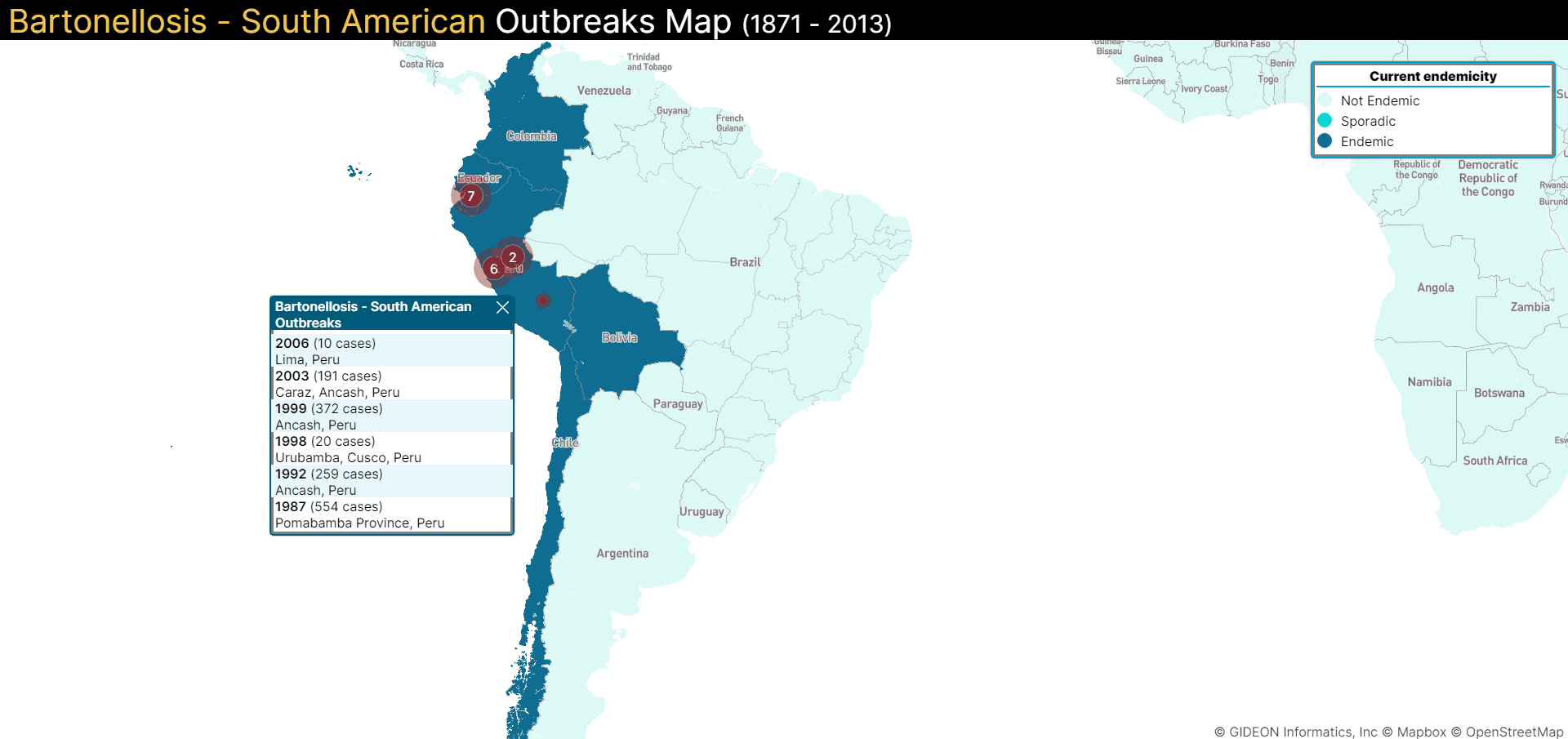
Oroya fever is quite deadly and, if untreated, increases the chance of mortality by 40% [15]. Peruvian warts, however, are not fatal [16]. Carrion’s disease is endemic to the mountainous regions of Colombia, Ecuador, and Peru and is considered a Neglected Tropical Disease (NTD) [13]. It is primarily found at elevations of 500 to 300 meters. Notable outbreaks include: 

  • 1871: The first outbreak of Carrion fever was recorded in Peru. 4,000-7,000 railroad workers died of Oroya fever. It began near La Oroya, a mining town where many railroad workers were working on a new railroad — a project that, unfortunately, brought them in contact with sand flies that were vectors of the disease. At the time, the origin of the disease was not known.
  • 1906: Another outbreak in Peru claimed the lives of 200 tunnel workers
  • 1959: 200 people died during an outbreak in Anco, a district in Peru
  • 2001 – 2005: An Oroya fever outbreak re-emerged in Ancash, Cajamarca, Amazonas, Piura, Cusco, La Libertad, Puno, and Ayacucho in South America [37]


How is it Spread?


Carrion’s disease is transmitted to humans when they are bitten by female sandflies (Lutzomyia verrucarum spp.) vectors carrying B. bacilliformis bacteria. These sandflies are usually found at high altitudes [7,17]. 

Biology


The first phase of Carrion’s disease is characterized by Oroya fever, which can be quite deadly. This is because red blood cells get infected when B. bacilliformis bacteria is released into the bloodstream, causing severe anemia and temporary immunosuppression. 

The second phase is an eruptive phase called Peruvian warts or verruga peruana, which can appear weeks or even two months after the Oroya fever phase. Note: The Peruvian warts phase is not always preceded by Oroya fever. The eruptions or warts that appear on the skin during this phase result from an infection of the endothelial cells and their pronounced proliferation [14].

Symptoms


The incubation period of B. bacilliformis is two to three weeks [18].  Symptoms include: 

  • Phase one: Oroya fever
    • Fever
    • Headache
    • Muscle aches
    • Abdominal pain
    • Severe anemia


  • Phase two: Peruvian warts (does not always have to be preceded by Oroya fever)
    • Red-to-purple vascular sores appear on the skin [19].

Diagnosis


Different methods are used to diagnose B. bacilliformis infection in humans. In the endemic regions, diagnosis is done simply based on symptoms. Peripheral blood smears are also used to detect the presence of bacteria. Another common method is blood culture. The blood samples are collected and stored at 4°C for a few weeks (usually two to six weeks) for the bacteria to grow. In recent times, PCR techniques have also been found to be effective in detecting infection [20]. 

Treatment


Antibiotics are a lifeline for people suffering from Carrion’s disease. Early intervention is key; delays or the absence of treatment can be disastrous [21]. 

The first phase of Carrion’s disease, Oroya fever, is treated using an antibiotic named Chloramphenicol (50 mg/kg/day for 3 days and then 25 mg/kg/day for two weeks or 14 days). For pregnant women, Chloramphenicol (50–100 mg/kg/day) and penicillin G (50,000–1,00,000 IU/kg/day) are given for two weeks. 

An antibiotic named Rifampicin (10 mg/kg/day) and Streptomycin (15–20 mg/kg/day) is used to treat Peruvian warts, the second phase of Carrion’s disease. The treatment can last for two to three weeks [12].

Prevention


  • Use insect repellents that are EPA-registered
  • Cover your body completely with long-sleeved shirts and trousers to avoid being bitten by sandflies
  • Since sand flies are active at dawn and dusk, staying indoors during these times is advisable [3]


Trench Fever


Trench fever is also known as five-day fever or quintan fever (quinta is five in Spanish). The bacteria Bartonella quintana is the causative agent for trench fever. As the name ‘five-day fever’ suggests, a characteristic symptom of this disease is a fever that lasts about 4 – 5 days. Trench fever is associated with homelessness, war, and unsanitary conditions where body lice thrive [22].

History


Trench fever was first identified in World War I when British forces stationed in France noticed an acute fever in soldiers — over hundreds of cases in a few months. Major J H P Graham is credited with discovering trench fever. Since laboratory tests at the time could not identify the source of the infection, the soldiers named the disease ‘trench fever.’ 

For a while, there was a huge debate in the medical community; experts disagreed about whether the disease was a new one or a new version of an older, known condition. One of the reasons why is that trench fever has a recurring fever as a symptom, similar to malaria. At the time, it was difficult to perform differential diagnoses accurately, given the inconclusive lab test results. Eventually, after some research, British authorities accepted trench fever as a new disease in 1916 [23].

In the same year, Captain T Strethill Wright proposed that body lice spread the disease after observing that the disease was prevalent in winters when mosquitoes and flies were not present [24]. It was later identified as a disease caused by the bacteria called Rickettsia (later renamed Bartonella quintana). Even after the war ended, there were reports of trench fever outbreaks in Europe, Asia, and North Africa [25]. 

Epidemiology


Studies show that homeless individuals and alcoholics are more likely to be infected by B. quintana. The disease is endemic to Russia and Eastern Europe. In more recent times, the disease is known as ‘urban trench fever’ and can be found in populations of homeless and alcoholic individuals. People who migrate to big cities in Europe and North America from these regions are also often B. quintana hosts [25]. Trench fever has a low mortality rate and is not as deadly as Oroya fever [26]. Risk factors for spread include overcrowded and unsanitary conditions, poor body hygiene, war, and homelessness. 

How is it Spread?


Humans are the primary hosts for trench fever. Trench fever is transmitted through the feces of a human body louse that contains B. quintana [7]. The louse, a small insect, can live in the clothing, and the waste (feces) can enter the skin in areas where it’s broken. 

Biology


Trench fever is caused by B. quintana which breeds in the intestine of a human body louse. When a louse bites the skin of its host, it excretes the bacteria in its feces. Eventually, the individual scratches the area due to the itching sensation that is caused. This leads to microabrasions on the skin that facilitate the transmission of the bacteria [7]. 

Symptoms

Quintana infection has an incubation period of 15 to 25 days [27]. The following are some of the common symptoms:

  • Fever
  • Dizziness
  • Bone pain in the lower legs, neck, and back
  • Skin lesions 
  • Inflammation in the eye [25,27]

Diagnosis


The pathogen that causes trench fever is quite difficult to diagnose. A normal bacterial culture can be done to diagnose the infection. But, it takes at least 21 days for the bacteria to grow. Serology, PCR, and RT-PCR are much more effective [22]. 

Treatment


Antibiotics such as gentamicin (3 mg/kg/day for two weeks) and doxycycline (200 mg/day for four weeks) are commonly used to treat patients with trench fever [12]. Other antibiotics, such as erythromycin, or azithromycin, are also used to treat other infections caused by B. quintana. The course of oral antibiotics is typically a four to six-week course of oral antibiotics [25].

Prevention

  • Keep surroundings clean to avoid exposure to human body lice
  • Avoid sharing clothes, towels, or beds with strangers
  • Keep your body clean by regularly having a shower
  • Wear clothes that have been washed properly with hot water and dry them on high heat. This kills lice and their eggs [28]


Endocarditis


Endocarditis refers to an inflammation of the inner lining of the heart chambers and valves. This condition can be caused by bacteria or other germs that enter the human body, spread through the blood, and attach themselves to areas of the heart that are already damaged, like heart valves. This type of endocarditis can cause severe damage to heart valves and can be lethal if not treated in time. Infection by several Bartonella species can lead to endocarditis, including B. quintana and B. henselae — the two major sources [29,30]. 

History


Bartonella was first discovered as a source of endocarditis in 1993 after researchers reported three cases. In 1995, Drancourt et al. published their findings of B.quintana-related endocarditis in three homeless men in the New England Journal of Medicine [31].  


Epidemiology


Since different species of Bartonella can cause this particular condition, endocarditis can be found across a wide geographical range, with a larger prevalence reported in Europe and Africa, particularly in southern countries [32]. In Europe, Bartonella endocarditis prevalence is:

  • 0% in Sweden 
  • 1.1% in the United Kingdom
  • 3% in France and Germany
  • 9.8% in Tunisia
  • 15.6% in Algeria [32]

How is it Spread?

More than nine different Bartonella species have been known to cause endocarditis, and the mode of infection and pathogenesis depends on the causative agent [7,17]. It can be transmitted through a cat scratch, by the human body louse, the bite of a sand fly, and other modes, depending on the Bartonella vector [7,17]. B. henselae can be transmitted through cat scratches [33]. B.quintana is transmitted through the human body louse, and when the bacteria travel in the blood to the heart, it can cause endocarditis. 

Biology


Bacterial endocarditis impairs the working of the heart valves, which means the heart has to work harder to pump blood out. This type of endocarditis can form bacterial clumps on the heart, which, if dislodged, can become a part of the bloodstream and potentially block blood vessels. Other organs may also get infected as the bacteria travels through the body [33]. 


Symptoms


As a result, the incubation period is estimated to be between one to four weeks [3,27]. Common symptoms include:

  • Fatigue
  • Muscle pain
  • Joint pain
  • Headache
  • Chills
  • Nausea
  • Vomiting [34].

Diagnosis

Endocarditis caused by Bartonella can be diagnosed through PCR, immunofluorescence assay, or a western blot assay [35].

Treatment

More than 90% of endocarditis patients infected by Bartonella will need heart valve surgery [35]. Apart from surgery, treatment for Bartonella endocarditis includes antibiotics and may last for up to six weeks. Patients are treated with gentamicin (3 mg/kg/day for two weeks) and doxycycline (200 mg/day for 6 weeks) [12]. For those allergic to penicillin, tetracycline or macrolide is used instead for a minimum of four weeks [29].

Prevention

  • Use an antibacterial soap to wash your body
  • Any cuts or grazes on the body will have to be washed as soon as you notice them
  • Consult a physician if there is any redness, swelling, or fluid discharge from the skin
  • Maintain good oral hygiene by brushing your teeth regularly [36]


References

[1] P. K. Mada, H. Zulfiqar, and A. S. J. Chandranesan, “Bartonellosis,” in StatPearls [Internet], StatPearls Publishing, 2022.

[2] K. Petríková et al., “Seroprevalence of Bartonella henselae and Bartonella quintana Infection and Impact of Related Risk Factors in People from Eastern Slovakia,” Pathogens, vol. 10, no. 10, p. 1261, 2021.

[3] CDC, “Bartonella henselae infection or cat scratch disease (CSD),” Centers for Disease Control and Prevention, 19-Jan-2022. [Online]

[4] Debré R, Lamy M, Jammet ML, Costil L, Mozziconacci P. La maladie des griffes de chat. Bull Mem Soc Med Hop Paris. 1950;66:76–9.

[5] Rolain, J. M., B. La Scola, Z. Liang, B. Davoust, and D. Raoult. 2001. Immunofluorescent detection of intraerythrocytic Bartonella henselae in naturally infected cats. J. Clin. Microbiol. 39:2978–2980.

[6] C. A. Nelson, A. R. Moore, A. E. Perea, and P. S. Mead, “Cat scratch disease: U.S. clinicians’ experience and knowledge,” Zoonoses Public Health, vol. 65, no. 1, pp. 67–73, 2018.

[7] F. Iannino, S. Salucci, A. Di Provvido, A. Paolini, and E. Ruggieri, “Bartonella infections in humans dogs and cats,” Vet. Ital., vol. 54, no. 1, pp. 63–72, 2018.

[8] A. M. McCord, S. I. Resto-Ruiz, and B. E. Anderson, “Autocrine Role for Interleukin-8 in Bartonella henselae -Induced Angiogenesis,” Infect. Immun., vol. 74, no. 9, pp. 5185–5190, 2006.

[9] C. Dehio, M. Meyer, J. Berger, H. Schwarz, and C. Lanz, “Interaction of Bartonella henselae with endothelial cells results in bacterial aggregation on the cell surface and the subsequent engulfment and internalization of the bacterial aggregate by a unique structure, the invasome,” Journal of Cell Science, vol. 110, no. 18, pp. 2141-2154, 1997.  

[10] C. A. Nelson, S. Saha, and P. S. Mead, “Cat-scratch disease in the United States, 2005–2013,” Emerg. Infect. Dis., vol. 22, no. 10, pp. 1741–1746, 2016.

[11] S. A. Klotz, V. Ianas, and S. P. Elliott, “Cat-scratch Disease,” Am. Fam. Physician, vol. 83, no. 2, pp. 152–155, 2011.

[12] E. Angelakis and D. Raoult, “Pathogenicity and treatment of Bartonella infections,” Int. J. Antimicrob. Agents, vol. 44, no. 1, pp. 16–25, 2014.

[13] M. Garcia-Quintanilla, A. A. Dichter, H. Guerra, and V. A. J. Kempf, “Carrion’s disease: more than a neglected disease,” Parasit. Vectors, vol. 12, no. 1, p. 141, 2019.

[14] C. Gomes and J. Ruiz, “Carrion’s disease: The sound of silence,” Clin. Microbiol. Rev., vol. 31, no. 1, 2018.

[15] J. V. Pai-Dhungat and F. Parikh, “Oroya fever and Daniel carrion -A fatal quest,” Japi.org. [Online]

[16] M. F. Minnick, B. E. Anderson, A. Lima, J. M. Battisti, P. G. Lawyer, and R. J. Birtles, “Oroya fever and verruga peruana: bartonellosis unique to South America,” PLoS Negl. Trop. Dis., vol. 8, no. 7, p. e2919, 2014.

[17] CDC, “Bartonella Infection: Transmission,” Centers for Disease Control and Prevention, 19-Dec-2019. [Online]. 

[18] L. G. Rubin, “Principles and Practice of Pediatric Infectious Disease (Third Edition),” W.B. Saunders, pp. 928-930, 2008.

[19] CDC, “Bartonella bacilliformis infection,” Centers for Disease Prevention and Control, 10-Jan-2022. [Online]. 

[20] J. del Valle Mendoza et al., “Diagnosis of Carrion’s disease by direct blood PCR in thin blood smear negative samples,” PLoS One, vol. 9, no. 3, p. e92283, 2014.

[21] M. J. Pons, C. Gomes, J. Del Valle-Mendoza, and J. Ruiz, “Carrion’s disease: More than a sand fly-vectored illness,” PLoS Pathog., vol. 12, no. 10, p. e1005863, 2016.

[22] O. Okorji, O. Olarewaju, and W. C. Pace, Trench Fever. StatPearls Publishing, 2021.

[23] R. L. Atenstaedt, “Trench fever: The British medical response in the great war,” J. R. Soc. Med., vol. 99, no. 11, pp. 564–568, 2006.

[24] T. S. Wright, “Some notes on trench fever,” Br. Med. J., vol. 2, no. 2900, pp. 136–138, 1916.

[25] M. E. Ohl and D. H. Spach, “Bartonella quintana and urban trench fever,” Clin. Infect. Dis., vol. 31, no. 1, pp. 131–135, 2000.

[26] “Facts about Bartonella quintana infection (‘trench fever’),” European Center for Disease Prevention and Control. [Online]

[27] S. Badiaga and P. Brouqui, “Human louse-transmitted infectious diseases,” Clin. Microbiol. Infect., vol. 18, no. 4, pp. 332–337, 2012.

[28] CDC, “Bartonella quintana infection,” Centers for Disease Prevention and Control, 14-Jan-2022. [Online]. 

[29] E. Angelakis and D. Raoult, “Pathogenicity and treatment of Bartonella infections,” Int. J. Antimicrob. Agents, vol. 44, no. 1, pp. 16–25, 2014.

[30] D. Raoult et al., “Outcome and treatment of Bartonella endocarditis,” Arch. Intern. Med., vol. 163, no. 2, pp. 226–230, 2003. 

[31] M. Drancourt et al., “Bartonella (Rochalimaea) quintana endocarditis in three homeless men,” N. Engl. J. Med., vol. 332, no. 7, pp. 419–423, 1995.

[32] P. Brouqui and D. Raoult, “New insight into the diagnosis of fastidious bacterial endocarditis,” FEMS Immunol. Med. Microbiol., vol. 47, no. 1, pp. 1–13, 2006.

[33] Chomel BB, Kasten RW, Williams C, Wey AC, Henn JB, Maggi R, Carrasco S, Mazet J, Boulouis HJ, Maillard R, Breitschwerdt EB. 2009. Bartonella endocarditis: a pathology shared by animal reservoirs and patients. Ann N Y Acad Sci 1166:120–126.

[34] A. Gupta and M. D. Mendez, “Endocarditis,” in StatPearls [Internet], StatPearls Publishing, 2022.

[35] S. Edouard, C. Nabet, H. Lepidi, P.-E. Fournier, and D. Raoult, “Bartonella, a common cause of endocarditis: a report on 106 cases and review,” J. Clin. Microbiol., vol. 53, no. 3, pp. 824–829, 2015.

[36] NHS, “Endocarditis – prevention,” NHS. [Online].  

[37] “Global Infectious Diseases and epidemiology network,” GIDEON, 28-Jun-2021. [Online]. Available: https://www.gideononline.com/. [Accessed: 14-Jun-2022].


Author
Chandana Balasubramanian

Chandana Balasubramanian is an experienced healthcare executive who writes on the intersection of healthcare and technology. She is the President of Global Insight Advisory Network, and has a Masters degree in Biomedical Engineering from the University of Wisconsin-Madison, USA.

Articles you won’t delete.
Delivered to your inbox weekly.